The Gastric Polyclonal Antibody from GeneBioSolution is a high-quality antibody developed for immunohistochemistry (IHC) applications on formalin-fixed, paraffin-embedded (FFPE) tissues. This antibody detects gastric mucosal antigens, making it a valuable marker for the identification of gastric epithelial cells and the evaluation of gastric carcinomas, metaplasia, and related lesions. This polyclonal antibody provides sensitive and broad-spectrum recognition of gastric antigens, delivering strong cytoplasmic staining in gastric mucosa with minimal background. It is commonly used as part of an antibody panel in the diagnosis of adenocarcinomas of the stomach and in differentiating gastric-origin tumors from other gastrointestinal or metastatic carcinomas. Beyond diagnostics, the Gastric Polyclonal Antibody is also useful in research applications for studying gastric differentiation, mucosal biology, and tumor progression. GeneBioSolution ensures rigorous quality control and validation across multiple tissue types, providing lot-to-lot consistency, reliable staining, and compatibility with both manual and automated IHC platforms. Clinical & Research Applications Identification of gastric mucosal differentiation Diagnostic aid in gastric adenocarcinomas and related malignancies Differentiation of primary gastric tumors from metastatic carcinomas Research into gastric mucosa, tumorigenesis, and gastrointestinal pathology
The Gastric Polyclonal Antibody is intended for use in immunohistochemistry (IHC) on formalin-fixed, paraffin-embedded (FFPE) tissues for the qualitative detection of gastric mucosal antigens. It serves as a diagnostic aid in identifying gastric epithelial differentiation and in the evaluation of gastric carcinomas, metaplastic changes, and related gastrointestinal lesions, when interpreted by a qualified pathologist in conjunction with appropriate controls.
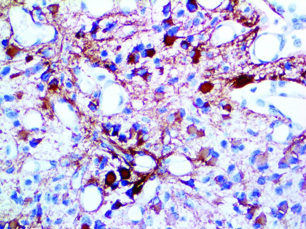
The Gastric Polyclonal Antibody recognizes antigens expressed in normal gastric mucosa and in a range of gastric-origin tumors. It produces strong cytoplasmic staining with high sensitivity, making it a valuable marker for differentiating primary gastric adenocarcinomas from other gastrointestinal or metastatic carcinomas. In addition to diagnostic pathology, it is widely used in research studies on gastric differentiation, mucosal biology, and cancer progression.
This antibody product may be used as the primary antibody in immunohistochemistry testing of formalin-fixed, paraffin-embedded tissue sections. In general, immunohistochemical (IHC) staining techniques allow for the visualization of antigens via the sequential application of a specific antibody to the antigen (primary antibody), a secondary antibody to the primary antibody (optional link antibody/probe), an enzyme complex and a chromogenic substrate with interposed washing steps. The enzymatic activation of the chromogen results in a visible reaction product at the antigen site. The specimen may then be counterstained, and cover slipped. Results are interpreted using a light microscope and aid in the differential diagnosis of pathophysiological processes, which may or may not be associated with a particular antigen.
Ployclonal
Human, Mouse, Rat, Cow, Pig, Rabbit, Chicken. Others not known.
Polyclonal
IgG1
Call for lot specific Ig concentration
The antibody recognizes antigenic epitopes associated with gastric mucosal glycoproteins and epithelial differentiation markers
Cytoplasmic staining in gastric mucosal epithelial cells. May also highlight areas of gastric metaplasia or carcinoma cells depending on expression pattern.
Tissues: Normal gastric mucosa, gastric adenocarcinoma, and metaplastic gastric lesions. Expected Staining: Strong, diffuse cytoplasmic positivity in gastric epithelial cells.
Immunohistochemistry (IHC): 30 min at RT. Staining of formalin-fixed tissues requires heating tissue sections in 10mM Tris with 1mM EDTA, pH 9.0, for 45 min at 95°C followed by cooling at RT for 20 minutes.
Buffer with protein carrier and preservative
