Fighting Cancer, One Slide at a Time

Product Cat. No.: GBS-003

For Research Use Only.

BCR/ABL gene fusion detection kit. Instructions Manual

Product Name

BCR/ABL gene fusion detection kit.

Package Specifications

10 Tests/box.

Intended Usage

This kit is mainly used for the detection of BCR/ABL gene fusion. The test samples are bone marrow cells suspected or diagnosed with leukemia patients (clinical or post-treatment patients) by clinical routine examination and used only for auxiliary diagnosis of the patient’s molecular typing.
Leukemia is a kind of malignant clonal disease of hematopoietic stem cells. Clonal leukemic cells proliferate and accumulate in bone marrow and other hematopoietic tissues, and infiltrate other non-hematopoietic tissues and organs, because of an uncontrolled proliferation, differentiation and apoptosis, and inhibition of the normal hematopoietic function. BCR/ABL gene is a common cytogenetic anomaly in patients with chronic myelocytic leukemia (CML). The BCR/ABL fusion gene can be found in 90% of CML patients, and the prognosis of the patients with BCR/ABL gene is poor.
This kit was validated against the BCR/ABL gene fusion detection performance only, and was not combined with the drug for clinical validation. This kit is only suitable for the detection of BCR/ABL gene fusion status, the test results are for clinical reference only and should not be used as the only basis for diagnosis. The clinician should make comprehensive judgment on the test results in combination with other clinical indicators.

Detection Principle

Fluorescence in situ hybridization is a technique for direct detection of specific nucleic acids in cells. According to the principle of complementary bases pairing, a specific probe is complementary to a target sequence within the cell. The probe and target sequence can be clearly observed under fluorescence microscope and under appropriate excitation light, due to the probe fluorescence.
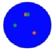
The kit uses orange fluorescein-labeled ABL probe and green fluorescein-labeled BCR probe. By in situ hybridization technique, the two probes bind to the target detection site. Normally (if BCR/ABL gene have not fused), two orange red signals and two green signals are shown under fluorescence microscope .When there is fusion, green and orange signals form by recombination a yellow fusion signal.
null

Product Main Components

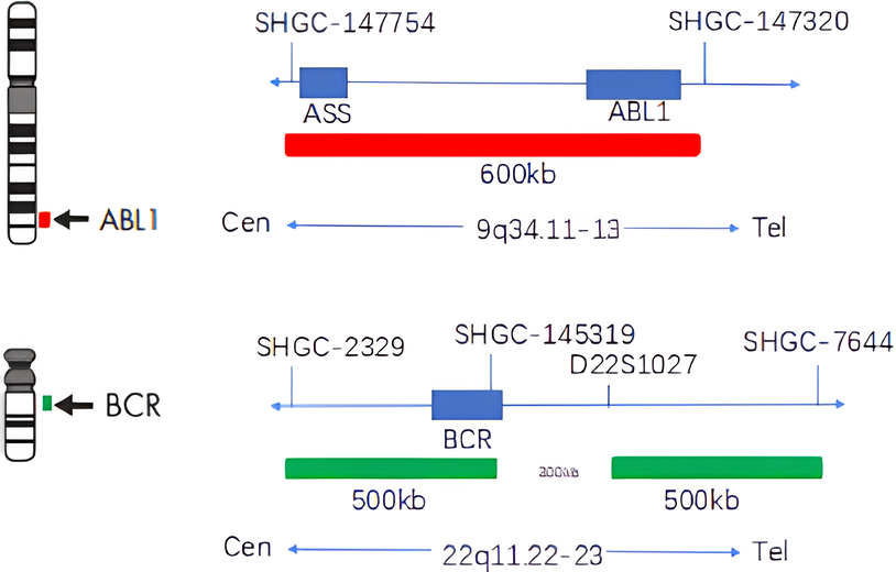
The kit consists of ABL orange probe and BCR green probe, as shown in Table 1. The reagents not provided in the kit are shown in Table 2.

Table 1: Kit composition

Component name Specifications Quantity Main components
BCR/ABL dual color probe 100μL/Tube 1 ABL orange probe, BCR green probe

Table 2: List of reagents not provided

Reagent name Purity Reagent name Purity
Sodium chloride Analytical purity AR NP-40 Analytical purity AR
Sodium citrate Analytical purity AR Xylene Analytical purity AR
Anhydrous ethanol Analytical purity AR Protease K ≥40 units/g

Storage conditions & Validity

Keep sealed away from light at -20oC±5oC. The product is valid for 12 months. Avoid unnecessary repeated freezing and thawing that should not exceed 10 times. After opening, within 24 hours for short-term preservation, keep sealed at 2~8oC in dark. For long-term preservation after opening, keep the lid sealed at -20oC±5oC away from light. See the label of the kit for the production date and expiration date.

Applicable Instruments

1. Fluorescence microscopy imaging system includes fluorescence microscope and filtersets. The kit islabeled with orange fluorescein, and the filter set compatible with the fluorescent labeled dye should be selected.
Orange fluorescence: The maximum excitation wavelength is 552nm and the maximum emission wavelength is 576nm.
Green fluorescence: The maximum excitation wavelength is 496nm and the maximum emission wavelength is 520nm.
Fluorescence microscopy imaging system should use a microscope with orange and green channels. For monochromatic channel microscope, image synthesis analysis results should be used.
2. Automatic hybridization instrument: Strict temperature uniformity is required, and the temperature difference should be ≤1oC.

Sample Requirements

1. Applicable specimen types: Fresh specimen that have not been fixed stored at 4°C for less than 24 hours; Cell suspensions after fixation stored at -20°C for less than 6 months; Prepared cell slides stored at -20°C for less than 1 month.
2. When specimen are stored at too high or too low a temperature (eg, frozen), the specimen will not be used for testing and should be discarded.
3. If the cell suspension is excessively volatile or contaminated during storage, the sample should be discarded.

Testing Method

1. Related reagents
The following reagents are required for the experiment but not provided in this kit

  • 20×SSC, pH 5.3±0.2

    Sodium chloride

    Sodium citrate

    176g

    88g

    Weigh 176g of sodium chloride and 88g of sodium citrate, dissolve in 800mL of deionized water, adjust the pH to 5.3±0.2 at room temperature, and complete to 1 L with deionized water. High-pressure steam sterilization, stored at 2~8oC, the solution shelf life is of 6 months. Discard if the reagent appears cloudy (turbid) or contaminated.

  • 2×SSC, pH 7.0±0.2
    Take 100mL of the above 20xSSC, dilute with 800mL deionized water, mix, adjust the pH to 7.0±0.2 at room temperature, complete to 1L with deionized water, stored at 2~8oC, the shelf life is of 6 months. Discard if the reagent appears cloudy (turbid) or contaminated.
  • Ethanol Solution: 70% ethanol, 85% ethanol
    Dilute 700ml, 850ml of ethanol with deionized water to 1L. The shelf life is of 6 months. Discard if the reagent appears cloudy (turbid) or contaminated.
  • 0.3% NP-40/0.4xSSC solution, pH 7.0 ~ 7.5

    NP-40

    0.6mL

    20xSSC

    4mL

    Take 0.6mL NP-40 and 4mL 20×SSC, add 150mL deionized water, mix, adjust the pH to 7.0 ~ 7.5 at room temperature, with deionized water complete to a volume of 200mL. Stored at 2~8°C, the shelf life is of 6 months. Discard if the reagent appears cloudy (turbid) or contaminated.

  • Fixation solution (methanol: glacial acetic acid = 3:1)
    Prepare a ready to use fixation solution by mixing thoroughly 30ml of methanol and 10ml of glacial acetic acid.
  • 0.075M KCl solution
    Weigh 2.8g of potassium chloride, dissolve in 400mL of deionized water and complete to 500mL with deionized water. Stored at room temperature, the solution shelf life is of 6 months. Discard if the reagent appears cloudy (turbid) or contaminated.
  • Diamidinyl phenylindole (DAPI) counterstain
    Use commercially available anti-quenching DAPI counterstain.

2. Sample collection and Slides preparation

  • Sample collection: Take anticoagulant cells sample.
  • Cells harvest: The cells sample is pipetted to the tip of a centrifuge tube and centrifuged at 1000rpm for 10 minutes to remove the supernatant.
  • Low permeability: Add 0.075mol/L KCL solution (6~8mL) pre-warmed at 37°C, mix with a pipette, and store in an incubator at 37°C for 20 to 30 minutes.
  • Pre-fixation: Add 2mL of 3:1 methanol, glacial acetic acid fixative solution and mix evenly. Centrifuge at 1000rpm for 10 minutes.
  • Fixation: Aspirate the supernatant; add freshly prepared 5mL of 3:1 methanol – glacial acetic acid fixative solution, mix evenly, fix for 10 minutes, centrifuge at 1000rpm for 10 minutes.
  • Repeat step 5 until cell pellets are precipitated, washed, and cleaned.
  • Cells suspension preparation: Pipet the supernatant and add the appropriate amount of fixative solution to prepare the appropriate cells suspension concentration.
  • Slides preparation: Pipet 3-5μl of cells suspension drop onto the slides, put at 56°C for 0.5-2 hours.
  • The prepared slide can be stored in a refrigerator at 4°C or in a refrigerator at -20°C for about 1 to 4 weeks.

3. Slides pretreatment

  • At room temperature, rinse the glass slides twice with SSC (pH 7.0) solution for 5min each time.
  • Place the glass slides in 70% ethanol, 85% ethanol and 100% ethanol and dry for 2 minutes.

4. Denaturation and Hybridization
The following operations should be performed in a darkroom.

  • Take out the probe put at room temperature for 5min. Mix and centrifuge briefly. Take 10μl droplet in the cell and drop in the hybridization zone, immediately cover 22mmx22mm glass slide area;spread evenly without bubbles the probe under the glass slide covered area and seal edges with rubber (edge sealing must be thorough to prevent dry film from affecting the test results during hybridization).
  • Place the glass slides in the hybridization instrument, denature at 88°C for 2 minutes (the hybridizer should be preheated to 88oC) and hybridize at 45°C for 2 to 16 hours.

5. Washing
The following operations should be performed in a darkroom.

  • Take out the hybridized glass slides, remove the rubber on the coverslip and immediately place the slides into 2xSSC for 5 seconds, and gently remove the coverslip. Place the glass slides in 2xSSC at room temperature for 1 min.
  • Remove and immerse the slidesin a 0.3% NP-40/0.4×SSC solution preheated at 68°C for 2 min. (Preparation of 0.3% NP-40/0.4xSSC: For 1L preparation, take 3mL NP-40 and 20mL 20xSSC, dissolve fully, mix well, and use 1M NaOH to adjust the pH to 7.2).
  • Immerse the glass slides in deionized water at 37oC for 1min, and dry naturally in the dark.

6. Counterstaining
The following operations should be performed in a darkroom.
Dip 10μL of DAPI counterstain into the hybridization area of the glass slides, immediately cover and then use the appropriate filter to observe the sections under the fluorescence microscope.
7. FISH results observation

  • Results observation method: Put the counterstained glass slide under the fluorescence microscope, and first put it under the low power objective lens (10x) to confirm the cell area under the microscope. Go to 40x under the objective lens, find a position where the cells are evenly distributed. Then, in the high-power objective (100x), observe the FISH results of nuclei. During microscopic examination, the continuous irradiation time of a single visual field under the green channel and red channel shall be controlled within 40 minutes.
  • Interpretable sample standard: The hybridization signal of the probe is bright and clear, the orange and green signals are easy to distinguish, the spontaneous fluorescence does not affect the signal count, and the number of countable cells is not less than 200.
  • Countable cell standard: The cell distribution is reasonable, there is no overlap, DAPI counterstaining is clear (i.e., the nuclear boundary is clear), and the number of orange, green signals or yellow signals formed by fusion in cells is ≥ 1.
  • Counting method: Randomly count 200 cells in each sample, count the number of orange, green, and yellow fusion signals in each nucleus, and calculate the ratio of cells showing abnormal cell signal mode (number of abnormal cells / number of counted cells) × 100%). Place the stained sections under a fluorescence microscope and confirm the cell area under a low magnification objective (10x). Under the 40x objective, ensure a uniform cell distribution. Then, under high magnification objectives (60x, 100x), observe nucleus size uniformity, nuclear boundary integrity, DAPI staining uniformity, absence of nuclei overlapping, and clear cell signals. Select at least 200 cells randomly and count the orange and green signals in the nuclei.

Positive judgment value

1. Common signal classification

Table 3: Dual-color for signal counting guide

Signal type

Diagram pattern

ABL

BCR

Cells results determination

Two orange red signals, two green
signals (2R2G)

Negative

One orange signal, one green signal,
two fusion signals (1R1G2F)

Positive

One orange signal, two green signals,
one fusion signal (1R2G1F)

Positive

One orange signal, one green signal,
one fusion signal (1R1G1F)

Positive

2. Threshold setting
Threshold should be set independently by each laboratory: Twenty human peripheral blood cell samples were randomly selected and processed according to sample processing requirements to prepare a deviant threshold reference slide. 200 cells were randomly counted in each reference slide. The number and percentage of cells that showed various types of positive cells were calculated, the mean and standard deviation of the statistical percentages were calculated.

  • Calculation of the total cells percentage with the 1R1G1F signal type, the mean and standard deviation of the statistical percentage, and the deviant threshold is set as the mean + 3 times the standard deviation, recorded as the threshold A.
  • Calculation of the total number cells percentage with other fusion signal types except 1R1G1F, the mean and standard deviation of the statistical percentage, and set the deviant threshold as the mean + 3 times the standard deviation, which is recorded as the threshold B.

The threshold A is 13.5% and the threshold B is 4%, can be used as a reference. Due to differences in sample processing methods and the subjective nature of signal counts, the laboratory thresholds will be different. Each laboratory should establish thresholds strictly in accordance with the standard process set by the threshold.

Interpretation of test results

1. FISH results determination
After counting 200 cells, the number and percentage of various types of positive cells were calculated respectively,

  • If the ratio of positive cells of 1r1g1f signal type > threshold a, it is judged as positive.
  • Except for 1r1g1f signal type, if the ratio of positive cells of other fusion signal types > threshold b, it is judged as positive.
  • If the ratio of positive cells of 1r1g1f signal type is less than threshold a, it is judged as negative.
  • Except for 1r1g1f signal type, if the ratio of positive cells of other fusion signal types < threshold b, it is interpreted as negative.
  • If the ratio of positive cells = threshold (a or b), increase the number of counts or analyze the whole film. If it is still less than or equal to the abnormal threshold, it will be interpreted as negative; otherwise, it will be interpreted as positive.
  • If the ratio of positive cells is within ±3 times the standard deviation of the mean value, the sample should be treated with caution.

2. Judgment of invalid experiment

  • If the number of cells available for probe analysis is less than 200, this test shall be judged as invalid.
  • If the fluorescence hybridization signal intensity or background available for analysis is not ideal or clear, which affects the judgment of results, this detection shall be judged as unreliable and treated as invalid experiment.
  • Common problems and treatment methods in the experiment
    The factors affecting the test results and treatment methodsin the experiment are shown in Table 4:

Table 4: FAQ and solutions

Question Possible cause Recommended solution

Too strong background

Inadequate washing after hybridization.

Improper use of filter sets.

Improper hybridization conditions.

Low washing temperature.

Ensure that the washing solution is prepared according to the instructions, ensure the correct pH value and temperature of the washing solution, remove the coverslip and repeat the washing steps.

Replace the appropriate filter setsfor observation and to weaken the background light.Ensure that the hybridization instrument temperature is 45°C.

Ensure that the hybridization instrument temperature is 45°C.

Ensure that the solution temperature is at the washing slides required temperature.

Too weak dye

Too weak dye.

Obsolete dye agent or excessive illumination

68°C 0.3%NP-40/0.4 × In SSc solution,shake for 10 ~ 20 seconds, remove the cover glass and soak for 2 minutes. Place the slide in deionized water at 37 °C and soak it for 1 minute. Dry the slide naturally in the dark and then re dye it.

Ensure that the dye agent is stored at -20°C and keep away from light. Make sure that the dye agent is valid.

No signal or weak signal

Specimen incomplete denaturation.

Improper pre-denaturation specimens’ preparation.

Probes and hybridization buffer improper mixture before usage.

Probe mixture on the slide dries too fast

Bubbles formation under coverslips during hybridization.

Inappropriate hybridization conditions.

Improper washing solution or washing conditions.

Probe or specimen slides inadequate storage.

Incorrect dye agent or too bright dye agent usage.

The selected filter sets is unsuitable for observation.

Ensure that the hybridization instrument temperature is at 88°C, and the hybridization instrument should be preheated at least 10min ahead of time.

Please refer to the above sample preparation related questions and answers.

Mix well the probe and the hybridization buffer, centrifuge briefly.

Wash the coverslip in the washing solution.

When covering the coverslip, cover the surface of the probe mix and squeeze gently to allow the bubbles to escape.

Ensure to observe time and temperature specified for the hybridization; do not leave gapsin the rubber seals; adjust hybridization time as appropriate.

Ensure that the washing solution is prepared according to the product specification; Check that the washing solution temperature reaches the in the washing step specified temperature; Assure that the thermometer and the pH meter are correctly calibrated.

Ensure that the probe is stored in dark at -20°C. Place the unhybridized slides dry at - 20°C for a long conservation or at room temperature for a short storage. Place the hybridized slides at -20°C and store in dark. The storage period should not exceed 6 months.

68°C 0.3%NP-40/0.4 × In SSc solution,shake for 10 ~ 20 seconds, remove the cover glass and soak for 2 minutes. Place the slide in deionized water at 37 °C and soak it for 1 minute. Dry the slide naturally in the dark and then re dye it.

Use the correct filter sets to observe the probe fluorescence. For details, please contact Gene Bio Solution, Technical Service Department.

Product Performance Index

  • Appearance: the outer package of the kit is complete without damage, and the marks are complete and clear; The liquid reagent shall be clearly marked without leakage.
  • Fluorescence signal intensity: after the probe is effectively hybridized with the karyotype reference, it will send out fluorescence signals that can be recognized by the naked eye under the fluorescence microscope.
  • Sensitivity: after the probe effectively hybridized with the karyotype reference, 100 chromosomes 22 of 50 cells in metaphase were analyzed, and at least 98 chromosomes 22 showed 1 green fluorescence signal; 100 chromosomes 9 of 50 cellsin metaphase were analyzed, and at least 98 chromosomes 9 showed an orange fluorescence signal.
  • Specificity: after the probe effectively hybridized with the karyotype reference, 100 chromosomes 22 of 50 cells in metaphase were analyzed, and at least 98 chromosomes 22 showed a specific green fluorescence signal in the long arm target area; 100 chromosomes 9 of 50 cells in metaphase were analyzed, and at least 98 chromosomes 9 showed a specific orange fluorescence signal in the long arm target region.
  • Coincidence rate of negative and positive: five negative reference materials(bone marrow cells of leukemia patients with BCR/ABL fusion gene negative clinical diagnosis) were detected and the fluorescence signals were analyzed. The results met the negative judgment criteria and were all judged to be negative. Five positive reference materials (bone marrow cells of leukemia patients with BCR/ABL fusion gene positive clinical diagnosis) were detected and the fluorescence signals were analyzed. The results met the positive judgment criteria and were all judged to be positive.
  • Precision: the precision of this kit within batch, between batches, within operation, between operation, during the day, and between operators / film readers is less than 5%.
  • Interfering substance: heparin sodium as anticoagulant will not interfere with the test results.
  • Clinical trial: among the 200 samples tested by this kit and the comparison reagent at the same time, the positive coincidence rate, negative coincidence rate and overall coincidence rate of this kit were 100%, and the kappa value was 1.00 (P < 0.001); In 1086 samples detected simultaneously by this kit and karyotype analysis, the positive coincidence rate of this kit was 96.39%, the negative coincidence rate was 99.13%, the overall coincidence rate was 98.71%, and the kappa value was 0.95 (P < 0.001).

Precautions

  • Please read this manual carefully before testing. The operator should undergo professional technical training. Signal counting personnel must be able to observe orange and green signals.
  • When testing clinical samples, the test will not provide any test results when the hybridization signal is difficult to count and the sample is not sufficient for repeated retests. If the amount of cells is not sufficient for analysis, the test will not provide test results.
  • The DAPI dye used in this experiment is potentially toxic or carcinogenic and should be handled in a fume hood. Wear masks and gloves to avoid direct contact.
  • The results of this kit will be affected by various factors of the sample itself, as well as restrictions such as enzyme digestion time, hybridization temperature and time, operating environment, and limitations of current molecular biology techniques, which may result in erroneous interpretation results. Users must understand the potential errors and accuracy limitations that may exist during the testing process.
  • All chemicals are potentially dangerous. Avoid direct contact. The used kits are clinical waste and should be properly disposed of.