Fighting Cancer, One Slide at a Time

Product Cat. No.: GBS-179-2

For Research Use Only.

EVI gene break apart probe reagent Instructions manual

Product Name

EVI gene break apart probe reagent

Package Specifications

10 Tests/box

Product Introduction

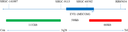
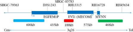
The kit is available in two pre-packages, Pre-Package 1 uses Orange and Green fluorescein to label the EVI1 probe, Pre-Package 2 uses Orange, Green and Aqua fluorescein to label the EVI1 probe, which can be conjugated to the target detection site by fluorescence in situ hybridization.The EVI1 probes are labeled with Orange (Green) and Cyan (Aqua) fluorescein.

Product Main Components

The kit consists of EVI1 dual color probe or EVI1 tri color probe as shown in Table 1.

Table 1: Kit composition

Cat# Component name Specifications Quantity Main components
GBS-179-1 EVI1 dual color probe 100μL/Tube 1 EVI1 Orange probe ; EVI1 Green probe
GBS-179-2 EVI1 tri color probe 100μL/Tube 1 EVI1 Orange probe ; EVI1 Green probe ; EVI1aqua probe

Storage conditions & Validity

Keep sealed away from light at -20°C±5°C. The product is valid for 12 months. Avoid unnecessary repeated freezing and thawing that should not exceed 10 times. After opening, within 24 hours for short-term preservation, keep sealed at 2-8°C in dark. For long-term preservation after opening, keep the lid sealed at -20°C±5°C away from light. The kit is transported below 0°C.

Applicable Instruments

Fluorescence microscopy imaging systems, including fluorescence microscopy and filter sets suitable for DAPI (367/452), Green (495/517), Aqua (423/480) and Orange (547/565).

Sample Requirements

  • Applicable specimen type: unfixed fresh bone marrow specimen (stored at 2-8°C for no more than 24 hours).
  • Sample collection: Take 1-3 mL of heparin sodium anticoagulated bone marrow cell sample.
  • Sample storage: After fixation, the cell suspension can be stored at -20±5°C for no more than 12 months. The prepared cell slides can be stored at -20±5°C for no more than 1 month. When the storage temperature of the specimen is too high or too low, or when the cell suspension is excessively volatile or contaminated during storage, the sample should not be used for testing.

Related Reagents

The following reagents are required for the experiment but not provided in this kit

  • 20×SSC, pH 5.3±0.2
    Weigh 176g of sodium chloride and 88g of sodium citrate, dissolve in 800mL of deionized water, adjust the pH to 5.3±0.2 at room temperature, and complete to 1 L with deionized water. High-pressure steam sterilization, stored at 2-8 °C, the solution shelf life is of 6 months. Discard if the reagent appears cloudy (turbid) or contaminated.
  • 2×SSC, pH 7.0±0.2
    Take 100mL of the above 20xSSC, dilute with 800mL deionized water, mix, adjust the pH to 7.0±0.2 at room temperature, complete to 1L with deionized water, stored at 2-8 °C, the shelf life is of 6 months. Discard if the reagent appears cloudy (turbid) or contaminated.
  • Ethanol Solution: 70% ethanol, 85% ethanol
    Dilute 700ml, 850ml of ethanol with deionized water to 1L. The shelf life is of 6 months. Discard if the reagent appears cloudy (turbid) or contaminated.
  • 0.3% NP-40/0.4xSSC solution, pH 7.0-7.5
    Take 0.6mL NP-40 and 4mL 20×SSC, add 150mL deionized water, mix, adjust the pH to 7.0-7.5 at room temperature, with deionized water complete to a volume of 200mL. Stored at 2-8 °C, the shelf life is of 6 months. Discard if the reagent appears cloudy (turbid) or contaminated.
  • Fixation solution (methanol: glacial acetic acid = 3:1): Prepare a ready-to-use fixation solution by mixing thoroughly 30mL of methanol and 10mL of glacial acetic acid.
  • 0.075M KCl solution: Weigh 2.8g of potassium chloride, dissolve in 400mL of deionized water, and complete to 500mL with deionized water. Store at room temperature. The solution has a shelf life of 6 months. Discard if the reagent appears cloudy (turbid) or contaminated.
  • Diamidinyl phenylindole (DAPI) counterstain: Use a commercially available anti-quenching DAPI counterstain.

Instructions

  • Sample collection and slide preparation
    • Sample collection: Take 1-3 mL of heparin sodium anticoagulated bone marrow cell sample.
    • Cell harvest: Pipet uncultured bone marrow cells or cultured bone marrow cell samples into a 15mL conical centrifuge tube, centrifuge at 500g for 5 minutes, carefully aspirate and discard the supernatant, and leave about 500μL of residual liquid to resuspend the cells.
    • Cell washing: Add 5mL of 1×PBS solution by pipetting to mix and resuspend the cell pellet, centrifuge at 500g for 5min, carefully aspirate and discard the supernatant, keep about 500μL of residual liquid to resuspend the cells; repeat once.
    • Cell permeation: Add 10mL hypotonic solution to each tube (pre-warmed at 37°C bath) and place at 37°C water bath hypotonic for 20min.
    • Cell pre-fixation: Add 1mL (10% volume) of fixative to the cell suspension after permeation to pre-fix the cells, gently pipette to mix, and immediately centrifuge at 500g for 5min, and remove the supernatant, keep about 500μL of residual liquid to resuspend the cells.
    • Cell fixation: Slowly add 10 mL of fixative to the cell suspension, put at room temperature for 10 min to fix the cells. Centrifuge at 500g for 5 min, and keep about 500μL of residual liquid to resuspend the cells; repeat once (the cells can also be fixed multiple times until the cells precipitate and wash out).
    • Preparation of cell suspension:After the last cell fixation and centrifugation, aspirate the supernatant and add an appropriate amount of fixative to prepare the cell suspension with the appropriate concentration.
  • Slide pretreatment procedure
    • Pretreatment: the slides were rinsed twice in 2×SSC solution at room temperature for 5min each time.
    • Dehydration: Place the cell drops in 70% ethanol, 85% ethanol, and 100% ethanol for 2 minutes each, then dry naturally.
  • Denaturation and Hybridization (Perform in a darkroom)
    • Take the probe at room temperature for 5 minutes. Briefly centrifuge manually (do not use vortex or shaker instrument). Take 10μl droplet in the cell and drop in the hybridization zone, immediately cover 22mmx22mm glass slide area; spread evenly without bubbles the probe under the glass slide covered area and seal edges with rubber (edge sealing must be thorough to prevent dry film from affecting the test results during hybridization).
    • Place the glass slide in the hybridization instrument, denature at 88°C for 2 minutes (the hybridizer should be preheated to 88°C) and hybridize at 45°C for 2 to 16 hours.
  • Washing (Perform in a darkroom)
    • Take out the hybridized glass slides, remove the rubber on the coverslip and immediately place the slides into 2xSSC for 5 seconds, and gently remove the coverslip.
    • Place the glass slides in 2xSSC at room temperature for 1 min.
    • Remove and immerse the slides in a 0.3% NP-40/0.4×SSC solution preheated at 68°C for 2 min.
    • Immerse the glass slides in deionized water at 37°C for 1min, and dry naturally in the dark.
  • Counterstaining (Perform in a darkroom)
    10μLDAPI compound dye is dropped in the hybridization area of the glass slide and immediately covered. The suitable filter is selected for glass slide observation under the fluorescence microscope.
  • FISH results observation
    Place the counterstained film under the fluorescence microscope, and first put it under the low-power objective lens (10x) confirm the cell area under the microscope; Go to 40x under the objective lens, find a position where the cells are evenly distributed; Then in the high- power objective (100x) the FISH results of nuclei are observed.

Common Signal Type Interpretation

EVI1 gene site 5 signal
EVI1 gene site 3 signal
dots

Negative:2 fusion

dots

Positive : 1 orange 1 green 1 fusion

dots

Positive : 2 fusion 1 green (suggests that the EVI1 breakpoint is located in the green labeled region)

dots

Positive : 2 fusion 1 orange (suggeststhat the EVI1 breakpoint is located in the orange labeled region)

EVI1 gene signal
EVI1 gene site 5 signal
EVI1 gene site 3 signal
dots

Negative:2 orange-green-aqua fusion

dots

Positive: 1 aqua; 1 orange-green fusion; 1 orange-green-aqua fusion

dots

Positive : 1 green; 1 orange-aqua fusion; 1 orange-green-aqua fusion (suggests possible EVI1 translocation)

dots

Positive : 1 orange-aqua fusion; 1 orange-green; 1 orange-green-aqua fusion

Precautions

1.The results of this kit will be affected by various factors of the sample itself, but also limited by hybridization temperature and time,operating environment and the limitations of current molecular biology technology, which may lead to wrong results.
2.Users must understand the potential errors and accuracy limitations that may exist in the detection process.
3.All chemicals are potentially dangerous. Avoid direct contact. Used kits are clinical waste and should be properly disposed off.