Fighting Cancer, One Slide at a Time

Product Cat. No.: GBS-045

For Research Use Only

1p/19q deletion probe reagent
Instructions Manual

Product Name

1p/19q deletion probe reagent.

Package Specifications

10 Tests/box

Product Introduction

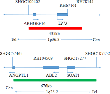
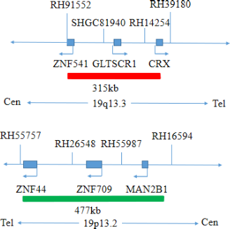
This kit uses orange fluorescent probes 1p36 and 19q13, green fluorescent probes 1q25 and 19p13 to bind 1p/19q probe to the target detection site by in situ hybridization.

Product Main Components

The kit consists of 1p36/1q25 dual-color probe and 19q13/19p13 dual-color probe as shown in Table 1.

Table 1: Kit composition

Component name Specifications Quantity Main components
1p36/1q25 dual color probe 100μL/Tube 1 1p36 orange probe ; 1q25 green probe
19q13/19p13 dual color probe 100μL/Tube 1 19q13 orange probe ; 19p13 green probe

Storage conditions & Validity

Keep sealed away from light at -20oC±5oC. The product is valid for 12 months. Avoid unnecessary repeated freezing and thawing thatshould not exceed 10 times. After opening, within 24 hours for short-term preservation, keep sealed at 2-8oC in dark. For long-term preservation after opening, keep the lid sealed at -20oC±5oC away from light. The kit should be shipped below 0°C.

Applicable Instruments

Fluorescence microscopy imaging system including fluorescence microscopy and filter sets suitable for DAPI (367/452), Green (495/517), and Orange (547/565).

Sample Requirements

  1. Applicable specimen types: Paraffin-embedded specimensfrom surgical excision or biopsy.
  2. The tissue should be fixed with 4% neutral formaldehyde solution within 1 hour after isolation. After tissue fixation, it is routinely dehydrated and embedded in paraffin.

Instructions

  1. Pre-hybridization treatment
    Baking: Slides heating at 80oC for 30min or 65oC for 2h or overnight.
    Dewaxing: According to the customer laboratory protocol (Commonly with Xylene for 15min).
    Hydration: Take out the slides and put them respectively into 100%, 85% and 70% EtOH at room temperature for 3 minutes each.
    Take out the slides, and immerse them in deionized water for 3 minutes. Remove the excess of water on the slides by air-drying.
    Permeation: Immerse the slides in deionized water at 100oC and boil continuously for 20-40 minutes (Conventional 20min). Remove the excess of water on the slides by air-drying.
    Digestion: Protease enzymic digestion at 37°C for 10-40 minutes. Mix the protease work buffer (50mmol HCl) and the 10x protease solution (Pepsin concentration 5%) in a proportion of 9:1 to prepare the enzymatic digestion solution.
    Washing: Wash with 2xSSC at room temperature for 5 minutes.
    Dehydration: Take out the slides and dehydrate in 70%, 85%, and 100% gradient ethanol at room temperature for 2 minutes each time. Remove the excess of EtOH solution on the slides by air-drying.
  2. Denaturation and Hybridization
    The following operations should be performed in a darkroom.

    1. Take the probe at static room temperature for 5 minutes. Briefly centrifuge manually (do not use vortex or shaker instrument). Take 10μl droplet in the cell and drop in the hybridization zone, immediately cover 22mmx22mm glass slide area; spread evenly without bubbles the probe under the glass slide covered area and seal edges with rubber (edge sealing must be thorough to prevent dry film from affecting the test results during hybridization).
    2. Place the glass slide in the hybridization instrument, denature at 85°C for 5 minutes (the hybridizer should be preheated to 85oC) and hybridize at 42°C for 2 to 16 hours.
  3. Washing
    The following operations should be performed in a darkroom.

    1. Take out the hybridized glass slides, remove the rubber on the coverslip and immediately immerse the slides in a 2xSSC solution for 5 seconds and remove the coverslip.
    2. Place the slides in a 2×SSC at room temperature for 1 min.
    3. Take out the slides and immerse in a preheated at 68°C 0.3% NP-40/0.4xSSC (Preparation of 0.3% NP-40/0.4xSSC: For 1L preparation, take 3mL NP-40 and 20mL 20xSSC, dissolve fully, mix well, and use 1M NaOH to adjust the pH to 7.2) solution and wash for 2min.
    4. Remove the slides and immerse in a 37°C preheated deionized water, wash for 1min and dry the slides naturally in the dark.
  4. Dyeing
    The following operations should be performed in a darkroom
    10μL DAPI compound dye is dropped in the hybridization area of the glass slide and immediately covered. The suitable filter is selected for glass slide observation under the fluorescence microscope.
  5. FISH results observation
    Place the counterstained film under the fluorescence microscope, and first put it under the low-power objective lens (10x) confirm the cell area under the microscope; Go to 40x under the objective lens, find a position where the cells are evenly distributed; Then in the high- ective (100x) the FISH results of nuclei are observed.

Common Signal Type Interpretation

1p36 and 19q13 signal
1q25 and 19p13 signal
dots

Negative: 2 Orange-red (2R) ; 2 Green (2G)

dots

Positive: 1 Orange-red (1R) ; 2 Green (2G)---- 1p36/1q25 point-out, 1p36 missing.

dots

Positive: 1 Orange-red (1R) ; 2 Green (2G)------ 19p13/19q13 point-out, 19q13 missing.

Precautions

  1. The results of this kit will be affected by various factors of the sample itself, but also limited by hybridization temperature and time, operating environment and the limitations of current molecular biology technology, which may lead to wrong results.
  2. Users must understand the potential errors and accuracy limitations that may exist in the detection process.
  3. All chemicals are potentially dangerous. Avoid direct contact. Used kits are clinical waste and should be properly disposed off.