Product Name
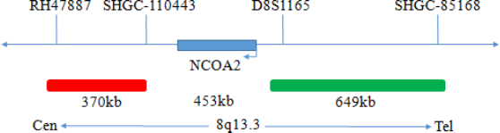
NCOA2(8q13) gene break apart probe reagent
Package Specifications
10 Tests/box
Intended Usage
This kit performs fluorescence in situ hybridization staining based on conventional staining, and provides auxiliary information for diagnosis for physicians. The test results are for clinical reference only and should not be used as the sole basis for clinical diagnosis. Clinicians should make comprehensive judgments on the test results based on factors such as the patient’s condition, drug indications, treatment response and other laboratory test indicators.
Detection Principle
Fluorescence in situ hybridization is a technique for directly observing specific nucleic acids in cells. According to the principle of base complementary pairing, the specific probe is complementary to the target sequence in the cell. Due to the fluorescence of the probe,the gene state of the hybrid probe and the target sequence can be clearly observed under the fluorescence microscope under the appropriate excitation light.
Product Main Components
This kit as shown in Table 1.
Storage conditions & Validity
Keep sealed away from light at -20oC± 5oC. The product is valid for 12 months.Avoid unnecessary repeated freezing and thawing that should not exceed 10 times. After opening, within 24 hours for short-term preservation, keep sealed at 2-8oC in dark. For long-term preservation after opening, keep the lid sealed at -20oC± 5oC away from light. The kit is transported under 0°C.
Applicable Instruments
Fluorescence microscopy imaging systems, including fluorescence microscopy and filter sets suitable for DAPI (367/452), Green (495/517),and Orange (547/565).
Sample requirements
Tissue:
1. Applicable specimen types: Paraffin-embedded specimens from surgical excision or biopsy.
2. The tissue should be fixed with 4% neutral formaldehyde solution within 1 hour after isolation. After tissue fixation, it is routinely dehydrated and embedded in paraffin.
Cell:
1. Take 1-3ml of heparin sodium anticoagulant bone marrow cells.
2. Sample preservation: Fresh bone marrow specimen without fixation (preserved at 2-8oC for no more than 24 hours). After fixation, the cell suspension can be preserved at -20±5oC for no more than 12 months; the prepared cell slide can be preserved at -20±5oC for no more than 1 month. When the storage temperature of the sample is too high or too low, the cell suspension is volatilized excessively or polluted,the sample cannot be used for detection.
Test method
1.Related reagents
- 20×SSC, pH 5.3±0.2
Weigh 176g of sodium chloride and 88g of sodium citrate, dissolve in 800mL of deionized water, adjust the pH to 5.3±0.2 at room temperature, and complete to 1 L with deionized water. High-pressure steam sterilization, stored at 2-8oC,the solution shelf life is of 6 months. Discard if the reagent appears cloudy (turbid) or contaminated.
- 2×SSC, pH 7.0±0.2
Take 100mL of the above 20xSSC, dilute with 800mL deionized water, mix, adjust the pH to 7.0±0.2 at room temperature, complete to 1L with deionized water, stored at 2-8oC, the shelf life is of 6 months. Discard if the reagent appears cloudy (turbid) or contaminated.
- Ethanol Solution: 70% ethanol, 85% ethanol
Dilute 700ml, 850ml of ethanol with deionized water to 1L. The shelf life is of 6 months. Discard if the reagent appears cloudy (turbid) or contaminated.
- 0.3% NP-40/0.4xSSC solution, pH 7.0-7.5
Take 0.6mL NP-40 and 4mL 20×SSC, add 150mL deionized water, mix, adjust the pH to 7.0-7.5 at room temperature, with deionized water complete to a volume of 200mL. Stored at 2-8oC, the shelf life is of 6 months. Discard if the reagent appears cloudy (turbid) or contaminated.
- Fixation solution (methanol: glacial acetic acid = 3:1)
Prepare a ready to use fixation solution by mixing thoroughly 30ml of methanol and 10ml of glacial acetic acid.
- 0.075M KCl solution
Weigh 2.8g of potassium chloride, dissolve in 400mL of deionized water and complete to 500mL with deionized water. Stored at room temperature, the solution shelf life is of 6 months. Discard if the reagent appears cloudy (turbid) or contaminated.
- Diamidinyl phenylindole (DAPI) counterstain
Use commercially available anti-quenching DAPI counterstain.
2. Sample processing before hybridization:
Tissue sample:
Baking: Slides heating at 80oC for 30min or 65oC for 2h or overnight.
Dewaxing: According to the customer laboratory protocol (Commonly with Xylene for 15min).
Hydration: Take out the slides and put them respectively into 100%, 85% and 70% EtOH at room temperature for 3 minutes each.Take out the slides, and immerse them in deionized water for 3 minutes. Remove the excess of water on the slides by air-drying.
Permeation: Immerse the slides in deionized water at 100oC and boil continuously for 20-40 minutes (Conventional 20min). Remove the excess of water on the slides by air-drying.
Digestion: Protease enzymic digestion at 37°C for 10-40 minutes. Mix the protease work buffer (50mmol HCl) and the 10x protease solution (Pepsin concentration 5%) in a proportion of 9:1 to prepare the enzymatic digestion solution.
Washing: Wash with 2xSSC at room temperature for 5 minutes.
Dehydration: Take out the slides and dehydrate in 70%, 85%, and 100% gradient ethanol at room temperature for 2 minutes each time.Remove the excess of EtOH solution on the slides by air-drying.
Cells sample:
- Sample collection: take 1-3ml of heparin sodium anticoagulant bone marrow cells.
- Cell harvesting: the uncultured marrow cells or the cultured marrow cell samples were aspirated to a 15mL centrifuged tube at the bottom of the tip, and centrifuged at 500g for 5min. The supernatant was carefully aspirated and discarded, leaving about 500μL of residual liquid to suspend the cells again.
- Cell washing: add 5ml of 1xPBS buffer solution, blow and mix up the heavy suspension cell precipitation, centrifugate 500g for 5min,carefully suck and discard the supernatant, and leave about 500μL of residual solution to heavy suspension cell; repeat once.
- Cell hypotonic: add 10ml of hypotonic solution to each tube (37oC warm bath in advance), and water bath at 37oC hypotonic for 20min.
- Cell pre fixation: add 1ml (10% volume) of fixed solution to the cell suspension after hypotonic treatment, gently blow and mix,centrifugate 500g immediately for 5min, remove the supernatant, and leave about 500μL of residual solution for cell suspension.
- Cell fixation: slowly add 10ml of the fixed solution to the cell suspension, leave it at room temperature for 10min to fix the cell, centrifugate 500g for 5min, and leave about 500μL of the residual solution to re suspend the cell; repeat once (or fix the cell several times until the cell is precipitated, washed and cleaned).
- Preparation of cell suspension: after the last centrifugation of cell fixation, the supernatant is sucked off, and a proper amount of fixed solution is added to make cell suspension with appropriate concentration.
- Preparation: take 3-10μL cell suspension drop to slide, aging at 56oC for 0.5h.
- The slides were rinsed twice in 2xSSC solution at room temperature for 5min each time.
- Dehydration: the cell drops were placed in 70% ethanol, 85% ethanol and 100% ethanol for 2 minutes respectively and then dried naturally.
3. Denaturation and Hybridization
The following operations need to be carried out in the darkroom.
Tissue samples:
- Take out the probe, let it stand at room temperature for 5min, turn it upside down with force, fully mix the probe, and then centrifuge briefly (vortex instrument oscillation is prohibited), take 10μL was dropped on the hybridization area of cell drops and immediately covered with 22mm×22mm cover glass, the probe shall be evenly expanded under the cover glass without bubbles, and the edge shall be sealed with rubber glue (the edge must be completely sealed to prevent the dry piece from affecting the test results during hybridization).
- Put the tissue sections on the hybridizer, and denature at 85°C for 5min (the hybridizer should be preheated to 85°C in advance), and hybridize at 42°C for 2-16h.
Cell sample:
- Take out the probe, leave it at room temperature for 5min, turn it upside down with force, mix it well, and then centrifuge it for a short time (no vortex instrument vibration). Take 10μL of it and drop it into the cell drop hybridization area, immediately cover the cover glass of 22mm × 22mm. The probe should be evenly expanded under the cover glass without bubbles, and seal the edge with rubber glue (the edge must be completely sealed to prevent the dry piece from affecting the test results in the hybridization process).
- The cell drops were placed on the hybridizer and denatured at 88oC for 2min (the hybridizer should be preheated to 88oC) and hybridized at 45oC for 2 to 16 hours.
4. Washing
The following operations need to be carried out in the darkroom.
- Carefully remove the sealing glue around the cover glass with tweezers to avoid sticking or moving the cover glass, immerse the sample in 2xSSC for about 5S, take it out, gently push a corner of the cover glass to the edge of the slide with tweezers, and gently remove the cover glass with tweezers.
- Place the sample at 2xSSC room temperature for 1 min.
- Take out the slides and immerse in a preheated at 68°C 0.3% NP 40/0.4xSSC (Preparation of 0.3% NP-40/0.4xSSC: For 1L preparation,take 3mL NP 40 and 20mL 20xSSC, dissolve fully, mix well, and use 1M NaOH to adjust the pH to 7.2) solution and wash for 2min.
- Take out the sample and immerse it in deionized water preheated at 37oC in advance for 1min; dry it naturally in the dark environment.
5. Counterstaining
The following operations should be performed in a darkroom
10μl DAPI compound dye is dropped in the hybridization area of the glass slide and immediately covered. The suitable filter is selected for glass slide observation under the fluorescence microscope.
6. FISH results observation
Place the counterstained film under the fluorescence microscope, and first put it under the low-power objective lens (10x) confirm the cell area under the microscope; Go to 40x under the objective lens, find a position where the cells are evenly distributed; Then in the high-power objective (100x) the FISH results of nuclei are observed.
Common signal type interpretation
NCOA2 gene site 3’signal
NCOA2 gene site 5’ signal
Positive : 1 orange 1 green 1 fusion
Precautions
1. Please read this manual carefully before testing. The testing personnel shall receive professional technical training, and the signal counting personnel must be able to observe and distinguish orange and green signals.
2. When testing clinical samples, when the hybridization signal counting is difficult and the sample is not enough to repeat the retest, or the cell volume is not enough for analysis, the test will not provide the test results.
3. DAPI counterstaining agent used in this experiment has potential toxicity or carcinogenicity, so it is necessary to operate in the fume hood,wear masks and gloves to avoid direct contact.
4. All chemicals are potentially dangerous. Avoid direct contact. Used kits are clinical waste and should be properly disposed off.