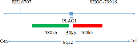
PLAG1(8q12) gene break apart probe reagent
10 Tests/box
This kit performs fluorescence in situ hybridization staining on the basis of conventional staining, and provides auxiliary information for diagnosis for physicians. The test results are for clinical reference only and should not be used as the sole basis for clinical diagnosis.Clinicians should make comprehensive judgments on the test results based on factors such as the patient’s condition, drug indications,treatment response and other laboratory test indicators.
The kit is based on fluorescence in situ hybridization technology. A nucleic acid probe islabeled with fluorescein. The target gene is detected with homologous complementary to the nucleic acid probe used. Both after denaturation, annealing and renaturation, the hybrid of the target gene and the nucleic acid probe can be formed, and the qualitative, quantitative or relative positioning analysis of the gene to be measured under the microscope can be performed by the fluorescence detection system.
The kit consists of PLAG1 dual-color probes, as shown in Table 1.
| Component name | Specifications | Quantity | Main components |
|---|---|---|---|
| PLAG1 dual color probe | 100μL/Tube | 1 | PLAG1 orange probe ; PLAG1 green probe, |
Keep sealed away from light at -20oC±5oC, and the validity period is 12 months. After the cover is opened, it can be sealed and stored in 2~8°C away from light within 24 hours. After the cover is opened, it should be sealed and stored in -20±5°C away from light for a long time. Transport with temperature below 0°C.
Fluorescence microscopy imaging systems, including fluorescence microscopy and filter sets suitable for DAPI (367/452), Green (495/517),and Orange (547/565).
1. Applicable specimen types: Paraffin-embedded specimens from surgical excision or biopsy.
2. The tissue should be fixed with 4% neutral formaldehyde solution within 1 hour after isolation. After tissue fixation, it is routinely dehydrated and embedded in paraffin.
Instructions
1. Hybridization pretreatment
Baking: Slides heating at 80oC for 30min or 65oC for 2h or overnight.
Dewaxing: According to the customer laboratory protocol (Commonly with Xylene for 15min).
Hydration: Take out the slides and put them respectively into 100%, 85% and 70% EtOH at room temperature for 3 minutes each.
Take out the slides, and immerse them in deionized water for 3 minutes. Remove the excess of water on the slides by air-drying.
Permeation: Immerse the slides in deionized water at 100oC and boil continuously for 20-40 minutes (Conventional 20min). Remove the excess of water on the slides by air-drying.
Digestion: Protease enzymic digestion at 37°C for 10-40 minutes. Mix the protease work buffer (50mmol HCl) and the 10x protease solution (Pepsin concentration 5%) in a proportion of 9:1 to prepare the enzymatic digestion solution.
Washing: Wash with 2xSSC at room temperature for 5 minutes.
Dehydration: Take out the slides and dehydrate in 70%, 85%, and 100% gradient ethanol at room temperature for 2 minutes each time.Remove the excess of EtOH solution on the slides by air-drying.
2. Denaturation and Hybridization
The following operations should be performed in a darkroom.
3. Washing
The following operations should be performed in a darkroom.
4. Counterstaining
The following operations should be performed in a darkroom.
Dip 10μL of DAPI counterstain into the hybridization area of the glass slides, immediately cover with a lid and place in dark for 10-20min,then use the appropriate filter to observe the sections under the fluorescence microscope.
5. FISH results observation
Place the counterstained film under the fluorescence microscope, and first put it under the low-power objective lens (10x) confirm the cell area under the microscope; Go to 40x under the objective lens, find a position where the cells are evenly distributed; Then in the high-power objective (100x) the FISH results of nuclei were observed.

Negative: 2 fusion

Positive: 1 orange 1 green 1 fusion
1. This product is only used for in vitro diagnosis.
2. Please read this manual carefully before testing. The testing personnel should receive professional technical training, and the signal counting personnel must be able to observe and identify tangerine and green signals.
3.When testing clinical samples, when the hybridization signal count is difficult and the sample is insufficient for repeated retesting, or the cell count is insufficient for analysis, the test will not provide test results.
4. The DAPI dye used in this experiment has potential toxicity or carcinogenicity, and should be operated in a fume hood, wearing masks and gloves to avoid direct contact.
5. All chemicals have potential hazards and should be avoided from direct contact. Used reagent kits are clinical waste and should be properly disposed off.
