Fighting Cancer, One Slide at a Time

Product Cat. No.: GBS-047

For Research Use Only.

PPARγ(3p25) gene break apart probe reagent Instructions Manual

Product Name

PPARγ(3p25) gene break apart probe reagent.

Package Specifications

10 Tests/box.

Intended use

The reagent carries out in situ hybridization staining on the basis of routine staining to provide doctors with auxiliary information for diagnosis. The test results are only for clinical reference and should not be used as the only basis for clinical diagnosis. Clinicians should comprehensively judge the test results in combination with the patient’s condition, drug indications, treatment response and other laboratory test indicators.

Detection principle

Fluorescence in situ hybridization is a technique for directly observing specific nucleic acids in cells. According to the principle of base complementary pairing, the specific probe is complementary to the target sequence in the cell. Due to the fluorescence of the probe, the gene state of the hybrid probe and the target sequence can be clearly observed under the fluorescence microscope under the appropriate excitation light.

Product Main Components

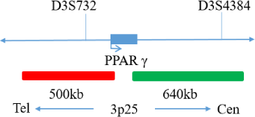
The kit consists of PPARγdual color probe as shown in Table 1.

Table 1: Kit composition

Component name Specifications Quantity Main components
PPARγ dual color probe 100μL/Tube 1 PPARγ Orange probe ; PPARγ Green probe

Storage conditions & Validity

Keep sealed away from light at -20°C±5°C. The product is valid for 12 months. Avoid unnecessary repeated freezing and thawing that should not exceed 10 times. After opening, within 24 hours for short-term preservation, keep sealed at 2-8°C in dark. For long-term preservation after opening, keep the lid sealed at -20°C±5°C away from light. The kit is transported below 0°C.

Applicable Instruments

Fluorescence microscopy imaging systems, including fluorescence microscopy and filter sets suitable for DAPI (367/452), Green (495/517), and Orange (547/565).

Sample Requirements

  1. Applicable specimen type: paraffin embedded specimen of surgical resection or biopsy tissue.
  2. The tissue should be fixed with 4% neutral formaldehyde fixation solution. After tissue fixation, it should be regularly dehydrated and embedded in paraffin.

Test method

  1. Sample processing before hybridization:
    Baking: Slides heating at 80°C for 30 min or 65°C for 2 h or overnight.Dewaxing: According to the customer laboratory protocol (commonly with Xylene for 15 min).
    Hydration: Take out the slides and put them respectively into 100%, 85%, and 70% EtOH at room temperature for 3 minutes each. Take out the slides and immerse them in deionized water for 3 minutes. Remove the excess water on the slides by air-drying.
    Permeation: Immerse the slides in deionized water at 100°C and boil continuously for 20-40 minutes (conventional 20 min). Remove the excess water on the slides by air-drying.
    Digestion: Protease enzymic digestion at 37°C for 10-40 minutes. Mix the protease work buffer (50 mmol HCl) and the 10x protease solution (Pepsin concentration 5%) in a proportion of 9:1 to prepare the enzymatic digestion solution.
    Washing: Wash with 2xSSC at room temperature for 5 minutes.
    Dehydration: Take out the slides and dehydrate in 70%, 85%, and 100% gradient ethanol at room temperature for 2 minutes each time. Remove the excess EtOH solution on the slides by air-drying.
  2. Denaturation and Hybridization
    The following operations should be performed in a darkroom.

    1. Take out the probe, put it at room temperature for 5 min. Mix and centrifuge briefly. Take a 10μl droplet in the cell and drop it in the hybridization zone, immediately cover a 22mm x 22mm glass slide area; spread evenly without bubbles under the glass slide covered area and seal edges with rubber (edge sealing must be thorough to prevent dry film from affecting the test results during hybridization).
    2. Place the glass slides in the hybridization instrument, denature at 85°C for 5 minutes (the hybridizer should be preheated to 85°C), and hybridize at 42°C for 2 to 16 hours.
  3. Washing
    The following operations should be performed in a darkroom.

    1. Take out the hybridized glass slides, remove the rubber on the coverslip, and immediately immerse the slides in a 2XSSC solution for 5 seconds and remove the coverslip.
    2. Place the slides in a 2×SSC at room temperature for 1 min.
    3. Take out the slides and immerse them in a preheated 68°C 0.3% NP-40/0.4xSSC solution (Preparation of 0.3% NP-40/0.4xSSC: For 1L preparation, take 3mL NP-40 and 20mL 20xSSC, dissolve fully, mix well, and use 1M NaOH to adjust the pH to 7.2) and wash for 2 min.
    4. Remove the slides and immerse them in 37°C preheated deionized water, wash for 1 min, and dry the slides naturally in the dark.
  4. Counterstaining
    The following operations should be performed in a darkroom.
    10μL DAPI compound dye is dropped in the hybridization area of the glass slide and immediately covered. The suitable filter is selected for glass slide observation under the fluorescence microscope.
  5. FISH results observation
    Put the counterstained cell drops under the fluorescence microscope, first under the low-power objective lens (10x) to confirm the cell area under the microscope.Go to 40x under the objective lens, find a position where the cells are evenly distributed.Then, in the high-power objective (100x), observe the FISH results of nuclei.

Common Signal Type Interpretation

PARTY gene site 5 signal
PARTY gene site 3 signal
dots

Negative : 2 fusion

dots

Positive : 1 orange 1 green 1 fusion

Precautions

  1. Please read this manual carefully before testing. The testing personnel shall receive professional technical training, and the signal counting personnel must be able to observe and distinguish orange and green signals.
  2. When testing clinical samples, when the hybridization signal counting is difficult and the sample is not enough to repeat the retest, or the cell volume is not enough for analysis, the test will not provide test results.
  3. DAPI counterstaining agent used in this experiment has potential toxicity or carcinogenicity, so it is necessary to operate in the fume hood, wear masks and gloves to avoid direct contact.
  4. All chemicals are potentially dangerous. Avoid direct contact. Used kits are clinical waste and should be properly disposed of.