NTRK1/NTRK2/NTRK3 Gene Break Apart Probe Detection Kit (Fluorescence In Situ Hybridization Method).
The reagent carries out in situ hybridization staining on the basis of routine staining to provide doctors with auxiliary information for diagnosis. The test results are only for clinical reference and should not be used as the only basis for clinical diagnosis. Clinicians should comprehensively judge the test results in combination with the patient’s condition, drug indications, treatment response and other laboratory test indicators.
Fluorescence in situ hybridization is a technique for directly observing specific nucleic acids in cells. According to the principle of base complementary pairing, the specific probe is complementary to the target sequence in the cell. Due to the fluorescence of the probe,the gene state of the hybrid probe and the target sequence can be clearly observed under the fluorescence microscope under the appropriate excitation light.
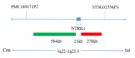
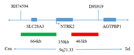
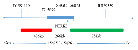
The kit consists of one of NTRK1, NTRK2 or NTRK3 dual color probe as shown in Table 1
| Component name | Cat.# | Specifications | Quantity | Main components |
|---|---|---|---|---|
| NTRK1dual color probe | GBS231-1 | 100μL/Tube | 1 | NTRK1 Orange probe, NTRK1 Green probe |
| NTRK2dual color probe | GBS-231-2 | 100μL/Tube | 1 | NTRK2 Orange probe, NTRK2 Green probe |
| NTRK3dual color probe | GBS-231-3 | 100μL/Tube | 1 | NTRK3 Orange probe, NTRK3 Green probe |
Keep sealed away from light at -20°C±5°C. The product is valid for 12 months. Avoid unnecessary repeated freezing and thawing that should not exceed 10 times. After opening, within 24 hours for short-term preservation, keep sealed at 2-8°C in dark. For long-term preservation after opening, keep the lid sealed at -20°C±5°C away from light. The kit is transported below 0°C.
Fluorescence microscopy imaging systems, including fluorescence microscopy and filter sets suitable for DAPI (367/452), Green (495/517),and Orange (547/565).
1. Applicable specimen types: Paraffin-embedded specimens for surgical resection or biopsy.
2. Tissue should be fixed with 4% neutral formaldehyde fixation solution, and the tissue should be fixed by conventional dehydration and paraffin embedding.
1. Sample Pretreatment
It is recommended to use GeneBio Solution’s “FISH Pretreatment Reagent ” (Cat.# GBS-003) for pretreatment.
2. Denaturation and Hybridization
The following operations should be performed in a darkroom.
3. Washing
The following operations should be performed in a darkroom.
4.Counterstaining
The following operations should be performed in a darkroom
10μl DAPI compound dye is dropped in the hybridization area of the glass slide and immediately covered. The suitable filter is selected for glass slide observation under the fluorescence microscope.
5. FISH results observation
Place the counterstained film under the fluorescence microscope, and first put it under the low-power objective lens (10x) Confirm the cell area under the microscope; Go to 40x under the objective lens, find a position where the cells are evenly distributed; Then in the highpower objective (100x) The fish results of nuclei are observed.

Negative: 2 fusion

Positive: 1 orange 1 green 1 fusion
Negative: 2 fusion
Positive: 1 orange 1 green 1 fusion
Negative: 2 fusion
Positive: 1 orange 1 green 1 fusion
1. Please read this manual carefully before testing. The testing personnel shall receive professional technical training. The signal counting personnel must be able to observe and distinguish orange red and green signals.
2. When testing clinical samples, if it is difficult to count the hybridization signals and the samples are not enough to repeat the retest, the test will not provide any test results. If the amount of cells is insufficient for analysis, again, the test will not provide test results.
3. The formamide and DAPI counterstaining agent used in this experiment have potential toxicity or carcinogenicity, so they need to be operated in the fume hood and wear masks and gloves to avoid direct contact.
4. The results of this kit will be affected by various factors of the sample itself, but also limited by enzyme digestion time, hybridization temperature and time, operating environment and limitations of current molecular biology technology, which may lead to wrong results. The user must understand the potential errors and accuracy limitations that may exist in the detection process.
5. All chemicals are potentially dangerous. Avoid direct contact. Used kits are clinical wastes and should be properly disposed of.
6. This product is for clinical diagnosis and scientific research.
