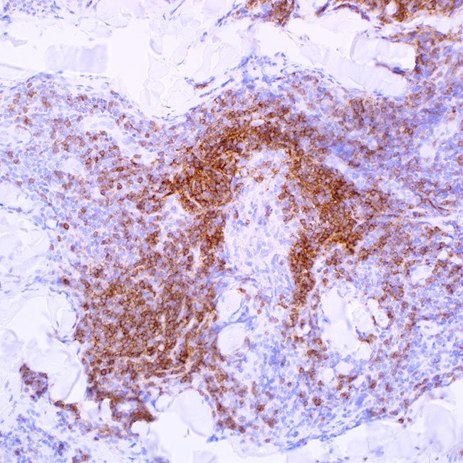
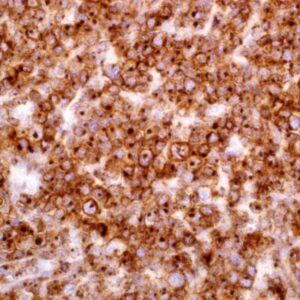
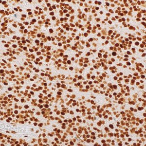
The monoclonal antibody against IgD interacts with immunoglobin D delta chains. Since the IgD antibody stains mantle zone cells in secondary follicles and mantle cells in primary follicles, immunohistochemical staining for IgD immunoglobulin heavy chain is frequently used to highlight the tonsil and nodal architecture in tonsil and lymph node. It has been discovered that IgD may be found in the surface/cytoplasm of neoplastic cells of common small B lymphoid cell lymphomas, such as small lymphocytic lymphoma, mantle cell lymphoma, marginal zone lymphoma (especially splenic marginal zone lymphoma), and follicular lymphoma.1-2 In subsets of instances (27% to 71.4%), IgD expression has been observed in L & P cells of nodular lymphocyte predominant Hodgkin lymphoma. Reed-Sternberg cells of classic Hodgkin lymphoma were found to be negative for IgD in studies. The IgD positive L & P cells are typically found in the extrafollicular region with a relatively T-cell-rich background. IgD expression is seldom observed in T-cell-rich B-cell lymphoma. IgD multiple myeloma is a rare bone marrow plasma cell dyscrasia that may be identified by the IgD antibody, particularly when a dry tap is encountered.